








Scientists Grow Human Cortical Tissue in Mouse Brains, Occupying About 92% of Their Cortex Volume
 ·
19.09.2026 15:36
·
3 min read
·
19.09.2026 15:36
·
3 min read
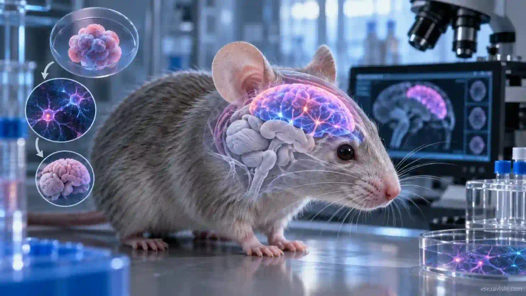
Stanford Medicine researchers have developed a new model to study human brain development: stem-cell-derived cortical organoids transplanted into newborn mice expanded substantially and integrated into the animals’ nervous systems. The study was published September 16 in Nature.
For the experiment, the team engineered “apallial mice.” Due to genetic modifications, these mice barely developed a neocortex or a large portion of the hippocampus. In adulthood, the animals’ own cortical tissue accounted for only about 2% of the normal volume.
Despite this, the mice were viable and appeared largely indistinguishable from normal mice, though researchers noted minor coordination and memory impairments. The vacated space allowed the human tissue to develop without intense competition from a rapidly growing rodent cortex.
Two-day-old mice were grafted with several cortical organoids containing roughly 100,000 cells each. Over the following months, the graft expanded nearly fivefold. After three months, human cells made up an average of 91.9% of the total cortical tissue volume in the studied animals.
The neurons did more than just survive: they established organized electrical activity, connected with the remaining mouse brain structures, and extended projections all the way down to the spinal cord.
Rare Human Neurons Emerged in the Tissue
One of the most notable findings was the emergence of von Economo neurons. These large, specialized cells are found in humans and select other social mammals, linked to brain regions involved in social behavior and decision-making.
Previously, such neurons were found primarily in postmortem human brain tissue and could not be generated in standard laboratory cultures. Researchers believe their appearance indicates that the living brain microenvironment enables human cells to undergo maturation stages that are difficult to replicate in a Petri dish.
The team also tested the model in a practical scenario. Following five hours of hypoxia, the human cortical tissue sustained noticeable damage, and the mice developed gait and balance issues. Normal and apallial mice tolerated the same level of oxygen deprivation significantly better. This makes the new platform potentially valuable for studying brain injuries linked to oxygen deprivation during pregnancy or labor.
The authors plan to use these “xenocortical mice” to research autism, epilepsy, schizophrenia, and neurodegenerative diseases, as well as to test potential drug candidates on human neurons within a functional nervous system.
However, describing these animals as “mice with human brains” would be inaccurate. As Stanford Medicine explains, human cells comprised the majority of the cortical tissue specifically, while the rest of the brain regions, the spinal cord, and the peripheral nervous system remained entirely mouse.