








Brain-IT Reconstructs Images People Have Seen from fMRI — an Hour of Data Is Enough to Adapt to a New Person
 ·
03.10.2026 15:49
·
3 min read
·
03.10.2026 15:49
·
3 min read
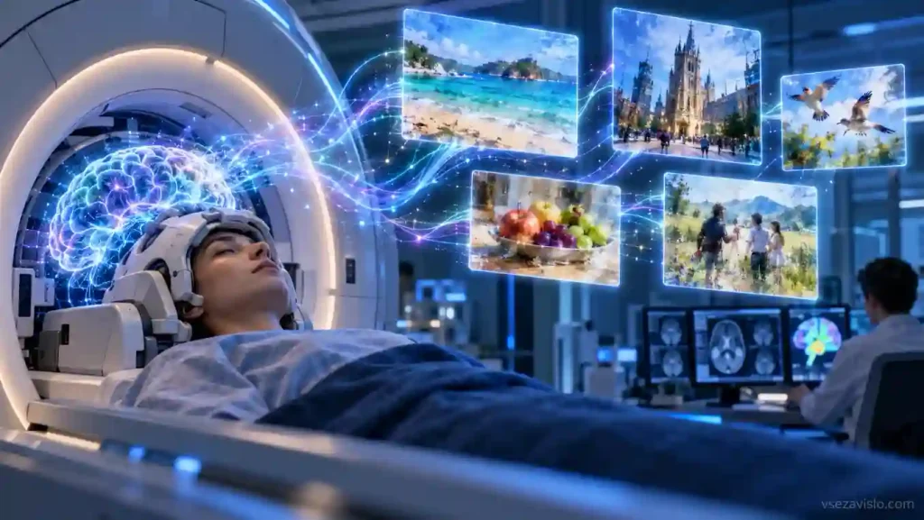
Researchers at the Weizmann Institute have developed Brain-IT, a system that reconstructs images from a person’s brain activity while they view them. The model analyzes functional MRI data and uses generative AI to try to recover the content of a scene, the arrangement of objects and some visual details. The work has been accepted to ICLR 2026, and the researchers published the source code on GitHub.
Despite the “mind reading” framing, Brain-IT currently handles a much narrower task. A person lies in an fMRI scanner and looks at a specific image, and the algorithm takes the brain activity recorded at that moment and tries to reconstruct what was seen. The system does not decode inner speech, memories or arbitrary thoughts.
One hour instead of dozens of hours of scanning
One of the main problems with such technologies is the need to train a model separately for each person. Different people’s brains are wired slightly differently, and collecting fMRI data takes a lot of time and requires expensive equipment.
In Brain-IT, the researchers group voxels — small volumetric elements of fMRI data — into functional clusters that perform similar tasks across different participants. As the project’s authors explain, the model works with two types of features at once. Semantic features help determine what is in the image, such as a face or food, while low-level features account for the overall structure and arrangement of objects.
This approach makes it possible to use knowledge gained from other people’s data. For a new participant, roughly one hour of individual fMRI recordings proved enough to approach the quality of previous methods trained on about 40 hours of data. In tests on the Natural Scenes Dataset, Brain-IT outperformed the compared approaches on seven of the eight metrics used.
An hour is not the absolute minimum, either: on the project page, the researchers show meaningful reconstructions even after 15 minutes of data. However, they use the one-hour result for a direct comparison with methods that required the full set of recordings.
This is a reconstruction, not a photograph from the brain
The resulting images are not exact copies of what was seen. Brain-IT can correctly reconstruct the type of scene, the main objects and their arrangement, but it may change the color, shape or specific details. The reason lies in the architecture itself: the system extracts visual features from fMRI, after which a diffusion model generates a suitable image rather than reading a ready-made frame from the brain.
This limitation is fundamental to understanding the technology. Based on the current results, Brain-IT shows how much information about visual perception can be extracted from brain activity rather than bringing a universal “mind scanner” closer.
The Weizmann Institute already names decoding sounds and, eventually, video as the next directions. The latter is noticeably harder because of fMRI’s low temporal resolution: a single measurement takes about two seconds, while video changes many times per second. So reconstructing dreams remains a possible future research direction rather than a feature of the existing Brain-IT.